Grants Funded
ASPS/PSF leadership is committed to continuing to provide high levels of investigator-initiated research support to ensure that plastic surgeons have the needed research resources to be pioneers and innovators in advancing the practice of medicine.
Research Abstracts
Search The PSF database to have easy access to full-text grant abstracts from past PSF-funded research projects 2003 to present. All abstracts are the work of the Principal Investigators and were retrieved from their PSF grant applications. Several different filters may be applied to locate abstracts specific to a particular focus area or PSF funding mechanism.
Predicting Tissue Necrosis in Mastectomy Flaps using Intra-Op Tissue Oximetry
Principal Investigator
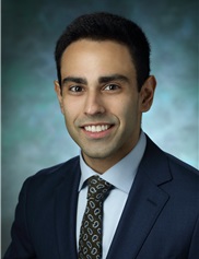
Nima Khavanin MD
Nima Khavanin MD
Year
2018
2018
Institution
Johns Hopkins Univ. School of Medicine
Johns Hopkins Univ. School of Medicine
Funding Mechanism
Scott Spear Innovation in Breast Reconstruction Fellowship
Scott Spear Innovation in Breast Reconstruction Fellowship
Focus Area
Breast (Cosmetic/Reconstructive), Technology Based
Breast (Cosmetic/Reconstructive), Technology Based
Abstract
Breast reconstruction has established itself as a pillar in the management of breast cancer, providing significant aesthetic and psychosocial benefits. Although oncologically safe, mastectomy interrupts blood flow to the remaining breast skin predisposing it to wound breakdown and skin loss. Particularly in immediate tissue expander reconstruction, poor perfusion of the skin flaps can lead to implant exposure and reconstructive failure with significant psychosocial harm, increased healthcare costs, and even delays in post-operative chemoradiation. Although an active area of research, no widely used technology currently exists to predict these issues intraoperatively. Surgeons are left to use subjective and inaccurate clinical findings to judge tissue viability. Recently the FDA approved a new handheld device, the Intra.Ox, (ViOptix, Inc., Fremont, CA) that uses near-infrared light to non-invasively measure tissue oxygenation intra-operatively. The device is easy-to-use, reusable, disposable, and early animal studies have demonstrated its ability to detect changes in tissue oxygen and predict the risk for tissue necrosis. Its clinical utility however, hinges upon its efficacy in a more relevant context in the management of human subjects. The overarching goals of this project are to develop evidence-based guidelines for intra-operative prediction and prevention of tissue necrosis. Specifically, in this study we aim to detect differences in tissue oxygenation at three time points, pre-operatively, after mastectomy, and after reconstruction. Additionally, we aim to correlate values obtained intra-operatively to a risk for tissue necrosis and use our data to identify thresholds at which tissue becomes at unviable. Finally, we will evaluate our ability to prevent complications by assessing changes in tissue oxygenation after the application of a known anti-necrosis agent, nitroglycerine paste, and correlate these measurements to tissue histology. We will take standardized measurements of tissue oxygenation in 120 women undergoing mastectomy and immediate breast reconstruction with a tissue expander. The surgical team will be blinded to these values, and the remainder of their management will proceed per the current standard of care. Additionally, we will perform translational studies in 24 rats using validated models for tissue necrosis to determine our ability to prevent this complication using intra-operative tissue oximetry.
Breast reconstruction has established itself as a pillar in the management of breast cancer, providing significant aesthetic and psychosocial benefits. Although oncologically safe, mastectomy interrupts blood flow to the remaining breast skin predisposing it to wound breakdown and skin loss. Particularly in immediate tissue expander reconstruction, poor perfusion of the skin flaps can lead to implant exposure and reconstructive failure with significant psychosocial harm, increased healthcare costs, and even delays in post-operative chemoradiation. Although an active area of research, no widely used technology currently exists to predict these issues intraoperatively. Surgeons are left to use subjective and inaccurate clinical findings to judge tissue viability. Recently the FDA approved a new handheld device, the Intra.Ox, (ViOptix, Inc., Fremont, CA) that uses near-infrared light to non-invasively measure tissue oxygenation intra-operatively. The device is easy-to-use, reusable, disposable, and early animal studies have demonstrated its ability to detect changes in tissue oxygen and predict the risk for tissue necrosis. Its clinical utility however, hinges upon its efficacy in a more relevant context in the management of human subjects. The overarching goals of this project are to develop evidence-based guidelines for intra-operative prediction and prevention of tissue necrosis. Specifically, in this study we aim to detect differences in tissue oxygenation at three time points, pre-operatively, after mastectomy, and after reconstruction. Additionally, we aim to correlate values obtained intra-operatively to a risk for tissue necrosis and use our data to identify thresholds at which tissue becomes at unviable. Finally, we will evaluate our ability to prevent complications by assessing changes in tissue oxygenation after the application of a known anti-necrosis agent, nitroglycerine paste, and correlate these measurements to tissue histology. We will take standardized measurements of tissue oxygenation in 120 women undergoing mastectomy and immediate breast reconstruction with a tissue expander. The surgical team will be blinded to these values, and the remainder of their management will proceed per the current standard of care. Additionally, we will perform translational studies in 24 rats using validated models for tissue necrosis to determine our ability to prevent this complication using intra-operative tissue oximetry.
Biography
 Both my research and clinical interests in breast cancer took root in college as I volunteered and did research in a major academic breast clinic. I was struck by how prevalent and devastating the disease could be, but also by the incredible bravery and resilience of the women who refused to be beaten by it. I immersed myself in the experience learning about the challenging journey of these patients and I realized that for many of these women, breast reconstruction represented an important milestone. It is the end of one chapter – the tumor is out – and the beginning of another where she can finally begin to feel like herself again.
I continued along this path in medical school, focusing more closely on plastic surgery and specifically breast reconstruction. I worked With Dr. John Kim at Northwestern Memorial authoring 14 peer reviewed research articles as well as a textbook chapter on the subject. Our work revolved around better understanding patient risk as well as application of various technologies in breast reconstruction. We applied the evidence we discovered to more objectively guide resource utilization and ultimately optimize patient outcomes.
Now a resident at the Johns Hopkins Hospital I am armed with the clinical experience and resources to make an even greater impact on the field. As breast cancer patients near the end of an already arduous journey, I hope to one day get them over this last hurdle unscathed - preventing complications altogether.