Grants Funded
ASPS/PSF leadership is committed to continuing to provide high levels of investigator-initiated research support to ensure that plastic surgeons have the needed research resources to be pioneers and innovators in advancing the practice of medicine.
Research Abstracts
Search The PSF database to have easy access to full-text grant abstracts from past PSF-funded research projects 2003 to present. All abstracts are the work of the Principal Investigators and were retrieved from their PSF grant applications. Several different filters may be applied to locate abstracts specific to a particular focus area or PSF funding mechanism.
Diagnosing Elevated Intracranial Pressure with Optical Coherence Tomography
Principal Investigator
Jordan Swanson MD, MSc
Jordan Swanson MD, MSc
Year
2018
2018
Institution
The Children's Hospital of Philadelphia
The Children's Hospital of Philadelphia
Funding Mechanism
AAPPS/PSF Research Grant
AAPPS/PSF Research Grant
Focus Area
Cranio/Maxillofacial/Head and Neck, Technology Based
Cranio/Maxillofacial/Head and Neck, Technology Based
Abstract
Elevated intracranial pressure has been implicated in 20-40% of patients presenting with craniosynostosis, and is increasingly suspected to play a role in neurocognitive impairment in a subset of these patients.1–4 Nonetheless, this relationship is not well understood, in particular due to limitations of conventional measures of intracranial pressure (ICP). Signs and symptoms of increased ICP, such as headaches and “thumbprinting” on CT scan are unreliable and have shown low sensitivity, particularly in young children.5 Traditional fundoscopy is subjective, and only detects about 1 in 5 patients with elevated ICP, often with late or severe findings.1,6–8 Direct ICP measurement with intracranial devices is invasive, prolonged, expensive, and associated with numerous complications.6,7 Optical coherence tomography (OCT) employs high-resolution laser interferometry to noninvasively quantify retinal thickness and has become a valuable tool for assessing the retina and optic nerve in older children and adults.9–12 Our center has completed a preliminary study suggesting that OCT can be potentially used as a noninvasive diagnostic tool of elevated ICP in children with craniosynostosis or hydrocephalus.13 Our aims are to prospectively: 1) validate through a large subject sample and potentially refine the OCT methodology developed in the previous pilot study among patients with intracranial pathology and normal control patients, 2) characterize patterns of elevated ICP among patients with craniosynostosis by diagnostic classification, and 3) determine clinical feasibility of deploying this OCT methodology at time of CT scan under sedation for infants and older children during clinic visits. We plan to achieve these aims by enrolling 185 subjects (100 craniosynostosis, 25 hydrocephalus, and 60 negative control). All subjects will be examined with pre-operative OCT, craniosynostosis and hydrocephalus patients will undergo pre-operative fundoscopy, and a subset of craniosynostosis and hydrocephalus patients will undergo direct intra-operative ICP measurement. A cohort of children within this sample will also undergo post-operative OCT evaluation without additional sedation. Negative control data will be used to establish normative values for five OCT parameters. Receiver operating characteristic (ROC) curves will be generated to examine the sensitivity and specificity of each OCT parameter. Data will be analyzed and reported for each of the three distinct aims.
Elevated intracranial pressure has been implicated in 20-40% of patients presenting with craniosynostosis, and is increasingly suspected to play a role in neurocognitive impairment in a subset of these patients.1–4 Nonetheless, this relationship is not well understood, in particular due to limitations of conventional measures of intracranial pressure (ICP). Signs and symptoms of increased ICP, such as headaches and “thumbprinting” on CT scan are unreliable and have shown low sensitivity, particularly in young children.5 Traditional fundoscopy is subjective, and only detects about 1 in 5 patients with elevated ICP, often with late or severe findings.1,6–8 Direct ICP measurement with intracranial devices is invasive, prolonged, expensive, and associated with numerous complications.6,7 Optical coherence tomography (OCT) employs high-resolution laser interferometry to noninvasively quantify retinal thickness and has become a valuable tool for assessing the retina and optic nerve in older children and adults.9–12 Our center has completed a preliminary study suggesting that OCT can be potentially used as a noninvasive diagnostic tool of elevated ICP in children with craniosynostosis or hydrocephalus.13 Our aims are to prospectively: 1) validate through a large subject sample and potentially refine the OCT methodology developed in the previous pilot study among patients with intracranial pathology and normal control patients, 2) characterize patterns of elevated ICP among patients with craniosynostosis by diagnostic classification, and 3) determine clinical feasibility of deploying this OCT methodology at time of CT scan under sedation for infants and older children during clinic visits. We plan to achieve these aims by enrolling 185 subjects (100 craniosynostosis, 25 hydrocephalus, and 60 negative control). All subjects will be examined with pre-operative OCT, craniosynostosis and hydrocephalus patients will undergo pre-operative fundoscopy, and a subset of craniosynostosis and hydrocephalus patients will undergo direct intra-operative ICP measurement. A cohort of children within this sample will also undergo post-operative OCT evaluation without additional sedation. Negative control data will be used to establish normative values for five OCT parameters. Receiver operating characteristic (ROC) curves will be generated to examine the sensitivity and specificity of each OCT parameter. Data will be analyzed and reported for each of the three distinct aims.
Biography
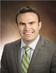
 Jordan Swanson, MD, MSc, is an attending surgeon in the Division of Plastic and Reconstructive Surgery at Children’s Hospital of Philadelphia who specializes in treatment of infants, children, adolescents and adults. Dr. Swanson focuses clinically on achieving optimal function and appearance of the face and head, and treats patients with cleft lip and palate, craniofacial, and other congenital, acquired and traumatic conditions. He specializes in cleft lip and palate repair, cranial vault and facial reconstruction, craniomaxillofacial distraction osteogenesis, rhinoplasty, otoplasty, and maxillo-mandibular (orthognathic) advancement, microsurgery, as well as minor pediatric plastic surgical procedures. In consultations and surgery, he brings attention to anticipating growth and development, striving to optimize treatment timing and procedure selection accordingly.
 Jordan Swanson, MD, MSc, is an attending surgeon in the Division of Plastic and Reconstructive Surgery at Children’s Hospital of Philadelphia who specializes in treatment of infants, children, adolescents and adults. Dr. Swanson focuses clinically on achieving optimal function and appearance of the face and head, and treats patients with cleft lip and palate, craniofacial, and other congenital, acquired and traumatic conditions. He specializes in cleft lip and palate repair, cranial vault and facial reconstruction, craniomaxillofacial distraction osteogenesis, rhinoplasty, otoplasty, and maxillo-mandibular (orthognathic) advancement, microsurgery, as well as minor pediatric plastic surgical procedures. In consultations and surgery, he brings attention to anticipating growth and development, striving to optimize treatment timing and procedure selection accordingly.