Grants Funded
ASPS/PSF leadership is committed to continuing to provide high levels of investigator-initiated research support to ensure that plastic surgeons have the needed research resources to be pioneers and innovators in advancing the practice of medicine.
Research Abstracts
Search The PSF database to have easy access to full-text grant abstracts from past PSF-funded research projects 2003 to present. All abstracts are the work of the Principal Investigators and were retrieved from their PSF grant applications. Several different filters may be applied to locate abstracts specific to a particular focus area or PSF funding mechanism.
A Novel Intramuscular Near Infrared Spectroscopy Probe for Free Flap Monitoring
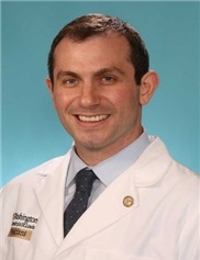
Mitchell Pet MD
2021
Washington University in St.Louis
ASRM/PSF Research Grant
Microsurgery, Technology Based
Impact Statement: Free tissue transfer is a critical tool which allows a reconstructive surgeon to move vascularized tissue (flaps) from areas of excess to areas of traumatic, oncologic, or congenital deficit. Skin and muscle flaps are commonly used for this purpose. It is critical to monitor blood flow to the flap, as blood clots within the flap vessels can cause catastrophic failure. Near-infrared spectroscopy (NIRS) has revolutionized flap monitoring, but current devices are only useful for skin flaps, leaving muscle flaps reliant upon inferior methods. Our novel intramuscular NIRS probe will bring this advanced monitoring technology to patients undergoing muscle flap reconstruction. This will elevate surgical success rates and improve the experience of the doctors, nurses, and patients.
Project Summary: As a result of trauma, cancer, or congenital differences, patients may be afflicted by critical deficits of tissue. Common situations include the absence of a breast after mastectomy or a shortage of tissue at the site of a fracture. Reconstructive microsurgery is used to replace what has been lost by transferring expendable tissue from another area of the body. This process is called free tissue transfer. While many types of tissue can be used, the two most common types of transferred tissue are skin and muscle. In order to transfer a large volume of tissue (or flap), it is necessary to provide blood flow to the tissue at its new location. This is accomplished by dissecting an artery and vein that supply the tissue mass, and attaching these to normal blood vessels near the tissue's destination. This is accomplished by direct suture of the artery and vein using a surgical microscope. Ongoing blood flow across these connections (or microvascular anastomoses) is critical for survival of the flap. Reconstructive failure can occur if a blood clot forms at one of the microvascular anastomoses. This stops blood flow and results in prompt tissue death if not surgically corrected within a few hours. Given the short window during which a flap can be rescued, careful monitoring and prompt detection are critical. The most advanced monitoring technology currently available is called near-infrared spectroscopy (NIRS). NIRS relies upon the detection of reflected light to measure the oxygen level present within the tissue. High oxygen levels indicate good blood flow, but low levels indicate the presence of a blood clot or other threat to the flap which requires surgical intervention. While this novel technology has revolutionized flap monitoring and improved patient outcomes substantially, it is unfortunately only available for flaps which include skin. This means that muscle flaps (which are favored for reconstruction of many traumatic injuries) continue to be monitored with older and less reliable technology. Our group has developed a new intramuscular NIRS sensor which directly addresses the unmet clinical need. This proposal is focused on rigorous pre-clinical testing and calibration of this novel sensor using a large animal model of flap malperfusion. It is our hope that by demonstrating excellent performance of this sensor for detecting troubled flaps in this animal model, we will be able justify translation of this technology into humans and help patients in ne
 I received my B.A. degree in 2007 from Dartmouth College, and my M.D. Degree in 2011 from Washington University in St. Louis. I went on to complete a residency in plastic and reconstructive surgery at the University of Washington Hospitals in from 2011 to 2017 and a hand/microsurgery fellowship at the Curtis National Hand Center. In September 2018, I returned to join the plastic and reconstructive surgery faculty at Washington University. I am the Associate Hand Surgery Fellowship director and my clinical practice involves both upper extremity surgery and complex microsurgical reconstruction from head to toe, and includes collaborative efforts with otolaryngology, dermatology, surgical oncology, and orthopedics.
My research career has been transformed by collaboration with Dr. Matthew MacEwan Washington University Department of Neurosurgery and Dr. John Rodgers from the Northwestern Querrey Simpson Institute for Bioelectronics. Using our combined experience with advanced biosensors, surgical practice/clinical needs, and translational research, we have been able to build a robust collaborative group which has already developed and tested several new technologies for flap monitoring.