Grants Funded
ASPS/PSF leadership is committed to continuing to provide high levels of investigator-initiated research support to ensure that plastic surgeons have the needed research resources to be pioneers and innovators in advancing the practice of medicine.
Research Abstracts
Search The PSF database to have easy access to full-text grant abstracts from past PSF-funded research projects 2003 to present. All abstracts are the work of the Principal Investigators and were retrieved from their PSF grant applications. Several different filters may be applied to locate abstracts specific to a particular focus area or PSF funding mechanism.
Preliminary validation of the Pediatric PainSCAN
Kristen Davidge MD
2022
Hospital for Sick Children (SickKids)
Pilot Research Grant
Peripheral Nerve, Other
Impact Statement: Our aim is to establish image-based artificial intelligence as a modality to predict and address key surgical problems, across surgical disciplines. Pre-operative surgical imaging contains unrecognized and unmeasured anatomic, structural, tissual, and relational features that can help surgeons quantify risk of surgical complications. Unlocking such data can power new domains of risk prediction to transform surgical care. The results of this project will be key for a future federally-funded proposal to evaluate using image-based feature analysis to optimize prediction for a wide variety of post-operative complications. These results will directly minimize complications, reduce surgical costs, and improve the patient experience.
Project Summary: Surgery is common and increasingly able to treat a greater scope of healthcare conditions. Surgical complications are prevalent, and affect between 3-16% of all patients undergoing inpatient procedures. They impact all stakeholders due to worsened health for patients, challenges for surgeons, and increased costs for the healthcare system. Predicting complications before surgery, therefore, is paramount. Current risk prediction tools are based exclusively on structured clinical data. However, 80% of healthcare data is unstructured and not easily extracted from Electronic Health Records. Our central hypothesis is that pre-operative surgical imaging in particular contains unrecognized and unmeasured features indicative of the pathophysiologic mechanisms that drive post-operative complications. Incisional hernia (IH), a common long-term complication of abdominal surgery, presents an ideal opportunity to test image-based risk prediction due to: 1) manifestation across surgical disciplines; 2) high costs and morbidity; 3) challenging prediction; and 4) simple clinical detectability. Pilot analysis of CT images for 100 abdominal surgery patients, matched 1:1 for IH development, has confirmed there are common anatomic phenotypes which are associated with IH formation. To further refine these phenotypes, our group aims to build upon pilot work by analyzing preoperative CT images for 100 additional matched patients. Following manual segmentation, these images will be assessed for morphometric, intensity-based, and texture-based features. Using our total 200 patient studies, we will employ the validated Optimal Biomarker (OBM) methodology to distill the most salient features that portend hernia risk. The 5-10 most potent OBMs will be incorporated into a model to predict IH formation. The final model will be assessed for accuracy of prediction and area under the curve. A multi-fold cross validation analysis will be performed using balanced and unbalanced techniques. Our trans-disciplinary approach to image-based risk analysis will ultimately lead to a more vigorous model for IH formation. This line of research serves as a paradigm for future work using routine pre-operative imaging to predict risk of any post-operative complication.
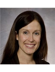 Dr. Davidge graduated from the University of Toronto Faculty of Medicine in 2005, and subsequently completed her residency training in Plastic & Reconstructive Surgery at the University of Toronto in 2012. During her residency, she completed a two-year Masters of Science degree in Clinical Epidemiology that was supported through local and national funding agencies. She pursued her fellowship training in hand, peripheral nerve, and microsurgery at the Washington University of St. Louis and in pediatric surgery at the Hospital for Sick Children. She then formally joined the Division of Plastic & Reconstructive Surgery at the Hospital for Sick Children in 2015. Her clinical practice centers on pediatric peripheral nerve injuries, congenital and acquired hand differences, and microsurgery. Her research program focuses on pain in brachial plexus birth injury, as well as function and patient-reported outcomes following nerve and hand surgery in children. She has been the recipient of numerous awards and grants during her career, has presented nationally and internationally on her research, and has authored many publications. Most recently, she was awarded of the George-Armstrong Peters Prize for outstanding productivity as a young research investigator from the Department of Surgery at the University of Toronto. She is also the current Program Chair for the American Society of Peripheral Nerve.