Grants Funded
Grant applicants for the 2024 cycle requested a total of nearly $3 million dollars. The PSF Study Section Subcommittees of Basic & Translational Research and Clinical Research evaluated more than 100 grant applications on the following topics:

The PSF awarded research grants totaling over $650,000 dollars to support more than 20 plastic surgery research proposals.
ASPS/PSF leadership is committed to continuing to provide high levels of investigator-initiated research support to ensure that plastic surgeons have the needed research resources to be pioneers and innovators in advancing the practice of medicine.
Research Abstracts
Search The PSF database to have easy access to full-text grant abstracts from past PSF-funded research projects 2003 to present. All abstracts are the work of the Principal Investigators and were retrieved from their PSF grant applications. Several different filters may be applied to locate abstracts specific to a particular focus area or PSF funding mechanism.
Migraine Surgery Candidate Selection with Magnetic Resonance Imaging
Salam Kassis MD
2022
Vanderbilt University Medical Center
Pilot Research Grant
Peripheral Nerve, Technology Based
Impact Statement: Our research provides a means to detect occipital nerve pathologies that contribute to some migraine headache disorders. This will reduce the time to diagnosis and treatment which will reduce the disability faced by these individuals. Additionally, a method of confirming the clinical diagnosis will improve the process of determining good surgical candidates who will respond to surgical decompression, thereby improving surgical outcomes. Furthermore, the impact of our study can be reasonably expected to extend beyond greater occipital nerve pathology because our extracranial nerve impingement MRI sequences can be modified and applied to several other extracranial nerves identified by nerve surgeons as being contributory to migraine headaches.
Project Summary: Migraine headaches (MH) are the 2nd greatest cause of years lived with disability (YLD) worldwide and affect 15% of the United States population. The past two decades have seen a rapid growth in research investigating an extracranial nerve contribution to MHs. Our study focuses on individuals with MHs that also have pain in the distribution of the greater occipital nerve and are scheduled for extracranial nerve decompression surgery to treat their MHs. We have previously developed extracranial nerve impingement magnetic resonance imaging (MRI) sequences that can track the greater occipital nerve from its origin to its point of entry into the subcutaneous tissue of the scalp. These sequences are sensitive enough to detect areas of compression which have been validated with intraoperative findings. Intraoperatively, the greater occipital nerve has been described to have several key areas in its course, near muscle fascia borders or the occipital artery, where visible compression is often evident as changes in color, pallor, and size. Edema or fibrosis may cause a paradoxical increase in size in the compressed area, but our MRI sequences can also detect anatomic variations where the occipital artery encircles the greater occipital nerve. However, without a quantitative measure of the intraneural architecture, the association of these findings with the headache symptomology remains uncertain. We therefore seek funding to pilot advanced MRI techniques that will add quantitative measurements of the intraneural architecture. Magnetic resonance (MR) diffusion tensor imaging (DTI) measurements include fractional anisotropy (FA) and apparent diffusion coefficient (ADC), and specific cut-off values of ADC and FA have been reported to diagnose and grade peripheral nerve compression pathologies. A second MRI technique, magnetization transfer ratio (MTR), allows quantification of myelin content which may provide useful prognostic insight for surgical candidates. We hypothesize that our DTI and MRT sequences will provide values of the greater occipital nerve that are significantly different from the nerve segment immediately proximal to the area of compression, and that these values will correlate with improvements in surgical outcome measures.
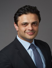
 Growing up in Lebanon instilled in me a desire to restore quality of life to people with nerve injuries and chronic pain. After completing my medical education and surgical internship at the American University of Beirut, I was intent to train in Plastic Surgery in the United States and to become the best reconstructive surgeon that I could be. My general surgery training continued at Yale before my dream of studying reconstructive surgery was realized at Vanderbilt University Medical Center.
During my plastic surgery fellowship, the faculty facilitated my building a multidisciplinary approach to migraine surgery that involved neurologists. Over my five years at Vanderbilt, I have worked on multiple nerve research projects. Our diffusion tensor imaging (DTI) research on nerves has been published in the Journal of Neurology, we have written two meta-analyses on DTI to diagnose compression neuropathies. We have a DOD grant to apply DTI to neuromas and are pursuing another DOD grant to use basic MRI sequences to establish the anatomy of the greater occipital nerve in a high quality clinical trial. The pilot grant for that DOD study provided the anatomic sequences needed for the present study, which will utilize advanced MRI techniques (DTI and magnetization transfer ratios) to quantitatively examine intraneural architectural for changes from nerve compression that we expect will indicate surgical management. Nerve surgery is a passion of mine that I will continue to pursue.